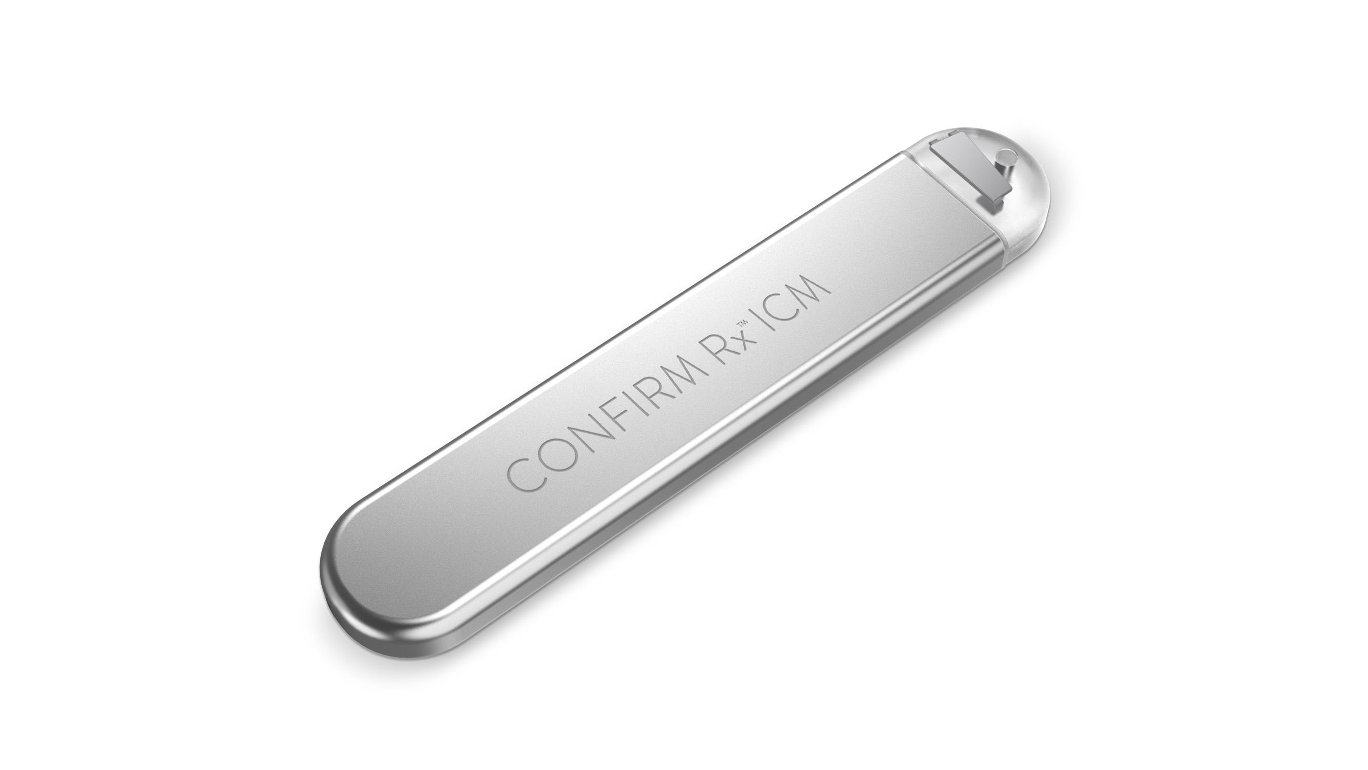
/cloudfront-us-east-1.images.arcpublishing.com/pmn/KAKXMK5Y2ZEYZHDMRX44366VJY.jpg)
Sampling at 1000 Hz only allows 1 channel vs 3 channels at sampling rates of 500 Hz or lower. A rate of 125 Hz does not sample frequently enough to allow sufficient fidelity, whereas higher frequencies may allow increased fidelity at the cost of battery life. This is because rhythm identification and filtering algorithms have all been optimized for 250 Hz. While capable of sampling at 125 Hz, 250 Hz, 500 Hz, and even 1000 Hz, the default sampling rate of all Preventice hardware is 250 Hz. In contrast, the Preventice (Houston, TX) Bodygaurdian Mini is preferentially placed in vertical orientation at the manubrium or alternatively in a horizontal (perpendicular) orientation at the same level based on body habitus.
Biotel heart monitor troubleshooting Patch#
While the BioTel MCOT samples at 250 Hz and provides 2 modified lead I channels, the iRhythm Zio Patch samples at 200 Hz and provides a single modified lead II channel. Both BioTel MCOT patch and iRhythm Zio Patch models are placed in the left parasternal region. Many monitoring platforms have also transitioned to a single hardware device for simplicity. Because polarity (unipolar vs bipolar), DBS programming (pulse amplitude and pulse width), proximity of the stimulator to recording device, and orientation of recording leads may all affect the degree of electromagnetic interference, it is likely that these factors influence the consistency and amplitude of the identified artifact. Only the stimulator being on or off determined artifact presence. We initially considered positional change as an etiology however, position did not affect presence of artifact during reproducibility testing. It is unclear why the artifact was only encountered 9% of the time. If any artifact is encountered on ECG, it is usually very low-amplitude, high-frequency noise, as exemplified in Figure 1 (notably leads I, III, AVL, and V 1). This would also explain why low-frequency artifact is not encountered on recordings obtained at a higher sampling rate such as the 12-lead ECG machine (sampling rate 500 Hz). This discrepancy, potentially compounded by proprietary signal processing algorithms and filters used by the monitor, likely results in the low-frequency aliasing artifact. In this case, the DBS stimulation frequency of 130 Hz (source) exceeds the Nyquist frequency by 5 Hz. The event monitor was placed in the left parasternal region per manufacturer recommendations and samples at a rate of 250 Hz, exhibiting a Nyquist frequency of 125 Hz (half the sampling rate). The DBS was programmed at 2.8 V at 60 ms pulse width and a stimulating frequency of 130 Hz in unipolar configuration.
Biotel heart monitor troubleshooting generator#
The DBS pulse generator was implanted in the right infraclavicular space and lead electrodes were implanted in the left subthalamic nucleus in our patient. Why are the DBS-related artifacts low-frequency “flutter wave” in this case instead of the anticipated high-frequency signals? The low-frequency artifact may at least be partially explained by aliasing. Despite visual resemblance to typical flutter, artifacts could not be ruled out. Initial review of the tracings suggested an apparent rate-controlled atrial flutter ( Figure 2A ). A 30-day event monitor (BioTel MCOT patch BioTelemetry Inc, Eagan, MN) surprisingly reported a 9% atrial flutter burden. However, he had no clinical tremor with his DBS (Activa 37601 Medtronic, Minneapolis, MN) and carbidopa-levodopa therapy, and no artifact was seen on the ECG obtained in our office. The ECG recordings obtained prior to cancellation of his colonoscopy were not available for review, but we initially considered the possibility of Parkinson-related tremor artifact superimposed upon an irregular rhythm owing to his premature atrial complexes being mistaken for atrial fibrillation. The patient had been asymptomatic from a cardiac standpoint, physical examination was within normal limits, and an echocardiogram revealed a structurally normal heart. A 12-lead electrocardiogram (ECG) obtained in the office revealed sinus rhythm with occasional premature atrial complexes ( Figure 1).

A 56-year-old man with history of Parkinson disease treated with a DBS was referred to electrophysiology for further evaluation for newly discovered atrial fibrillation on telemetry at the time of a routine preventative colonoscopy.


 0 kommentar(er)
